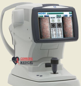
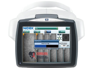
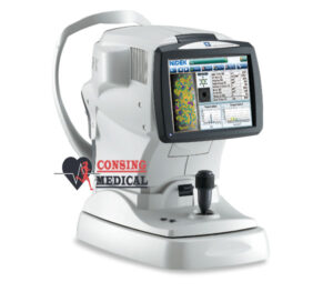
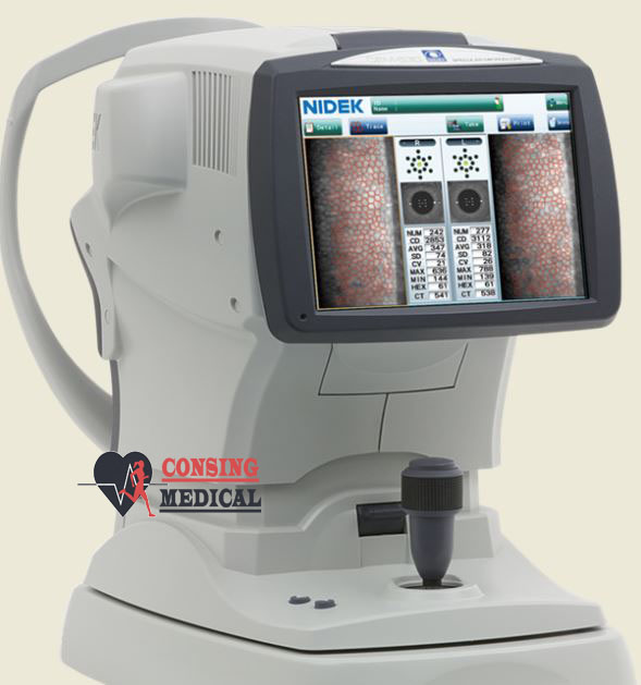
Nidek CEM-530 Specular Microscope

Nidek
The analysis results with graphic and color-coded cell images helps the clinician to rapidly and effectively evaluate the endothelial cell layer.
In addition to conventional central and peripheral specular microscopy, the nidek CEM-530 includes a unique function to capture paracentral images.
Paracentral specular microscopy
Faster measurements and two-second auto analysis
Comprehensive analysis
Advanced manual analysis functions
Easy operation
Additional features with CEM Viewer for NAVIS-EX
Combination of auto and manual analyses
All three manual analysis methods can be performed on the same image and also on auto-analyzed images. The versatility of combining automated and manual analyses allows analysis of the range of pathology in a comprehensive practice.

Auto tracking and auto alignment functions provide more accurate measurements increasing patient and operator comfort and efficiency.
These functions allow easy follow-up and reduce variations between examiners, resulting in well-aligned follow up exams.


Reviews
There are no reviews yet.